Pain Finder
Find the muscle behind the pain.
Tap the area that hurts. The muscles that refer pain to that area are shown.
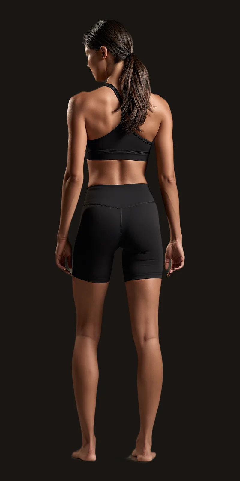
Tap a region
Head & Neck
Six referral patterns are shown here.
The Muscle Library lists all seventeen patterns.
Each muscle shows where the pain is felt, what causes it, and how patients describe it.
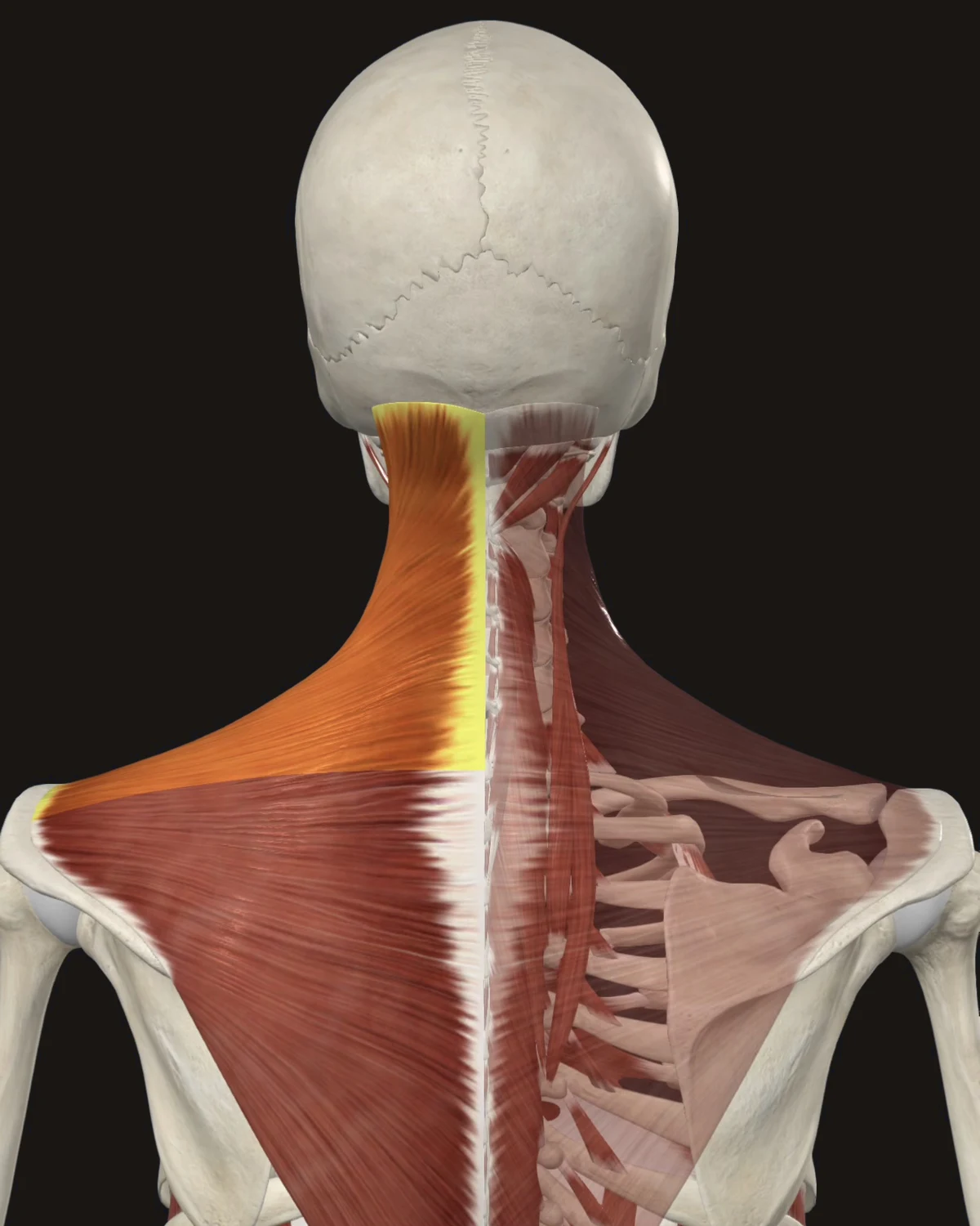
Where the pain is felt: Up the side of the neck to the base of the skull, wrapping to the temple and behind the eye. The mapped pattern is the classic “tension headache” path.
Common causes: Desk work with unsupported arms, phone held between ear and shoulder, heavy bags on one shoulder, stress-held shoulders, sleeping without neck support.
Patients say: “My shoulders live up by my ears.” A pulling ache up the neck by end of day. Headaches that start in the shoulders.
When to get evaluated: A sudden, severe headache unlike any before, or headache with fever, stiff neck, confusion, or vision change, is a medical evaluation, not a muscle pattern.
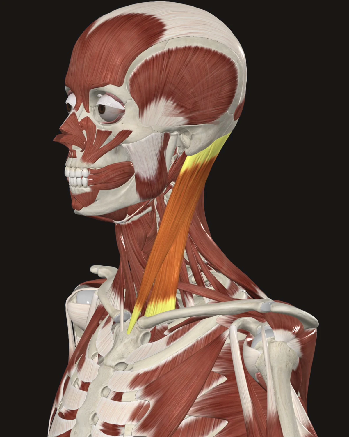
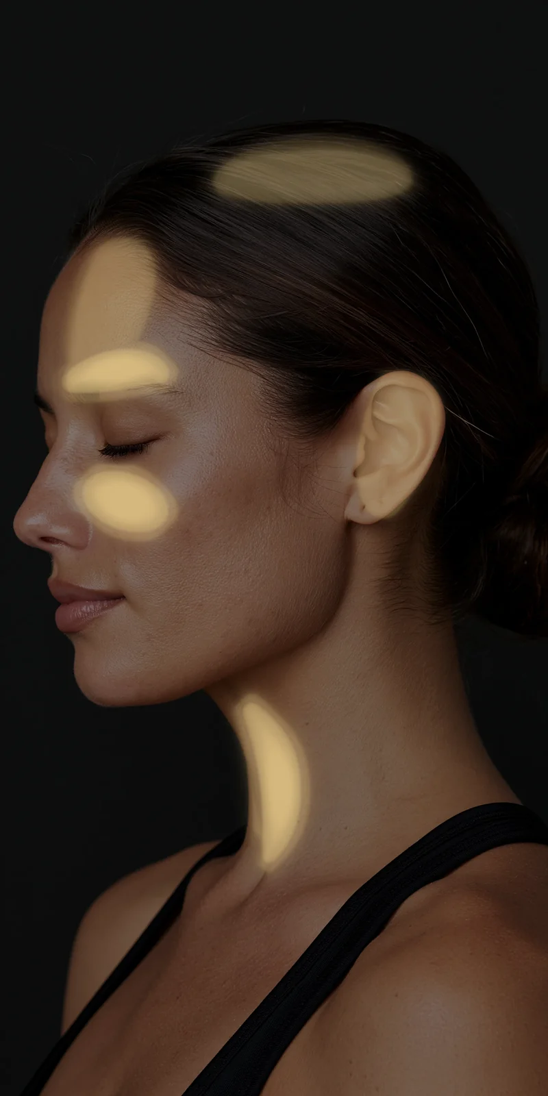
Where the pain is felt: The forehead, around and behind the eye, the ear, and the cheek. The muscle itself sits at the front of the neck, yet the pain shows up in the face and head.
Common causes: Forward-head posture, long screen sessions, painting ceilings or overhead work, whiplash, sleeping on the stomach with the head turned.
Patients say: Headache over the eye. Ear fullness or face pain with a normal ear exam. Dizziness when turning the head quickly.
When to get evaluated: New dizziness with hearing loss, slurred speech, facial droop, or loss of balance needs medical evaluation first.
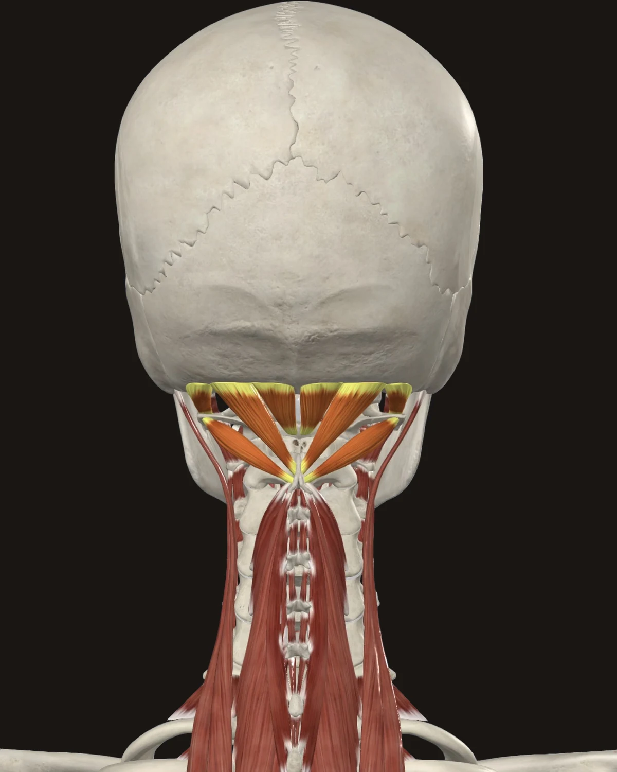
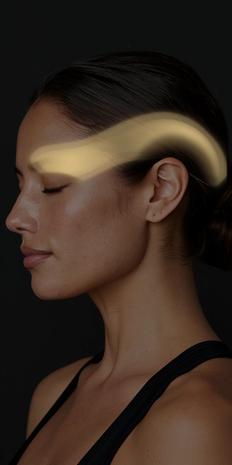
Where the pain is felt: A band from the base of the skull wrapping over the head to the eye. Described in the maps as pain inside the head rather than on it.
Common causes: Looking down at phones and laptops, reading glasses set too low, long driving, grinding through close-focus work.
Patients say: “It feels like a headband of pressure.” Ache at the skull base that climbs. Worse after a day of screens.
When to get evaluated: Headache that wakes you from sleep, worsens lying down, or arrives with nausea and vision change gets evaluated.
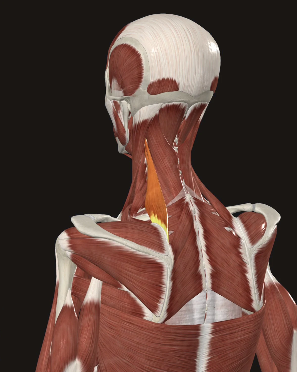
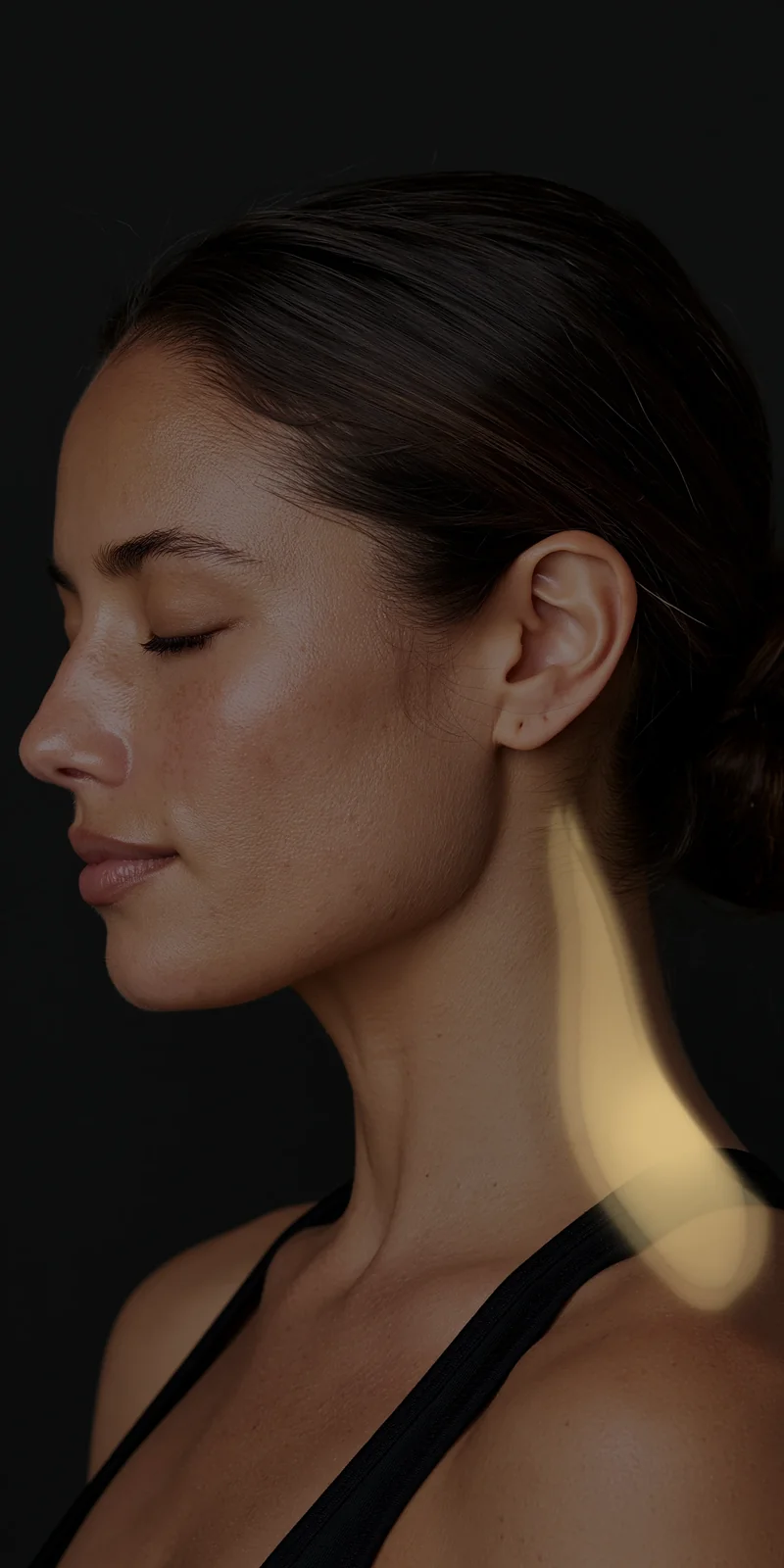
Where the pain is felt: The angle where the neck meets the shoulder, with a stiff line up the side of the neck. The signature restriction: turning the head to the same side.
Common causes: Sleeping with the neck bent, long phone calls, monitors set off-center, backpacks and shoulder bags, sustained stress posture.
Patients say: “I can’t check my blind spot.” Waking with a stiff neck. Pain at the exact corner of the neck and shoulder.
When to get evaluated: Neck stiffness with fever, or stiffness after an accident, is evaluated before any needling plan.
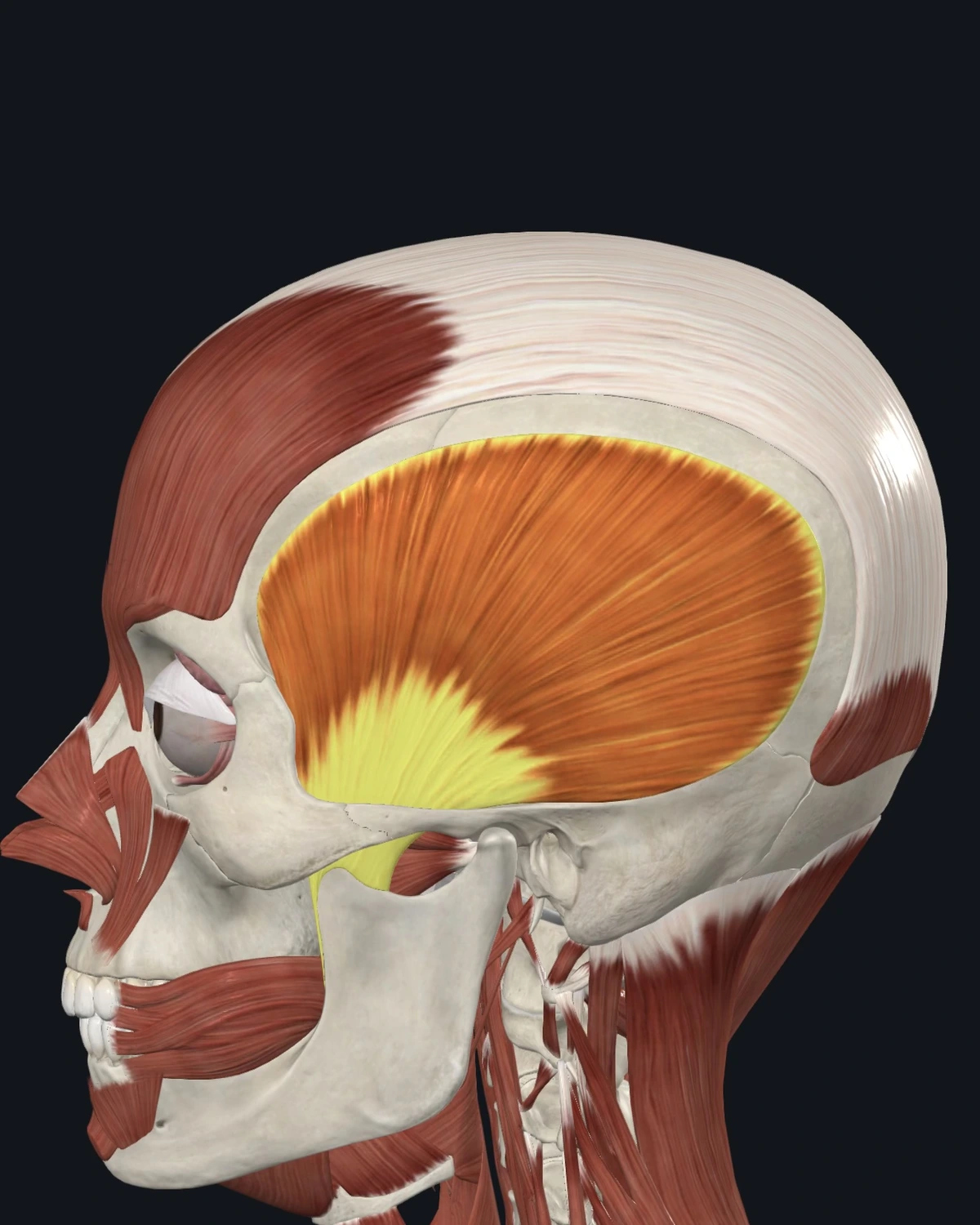
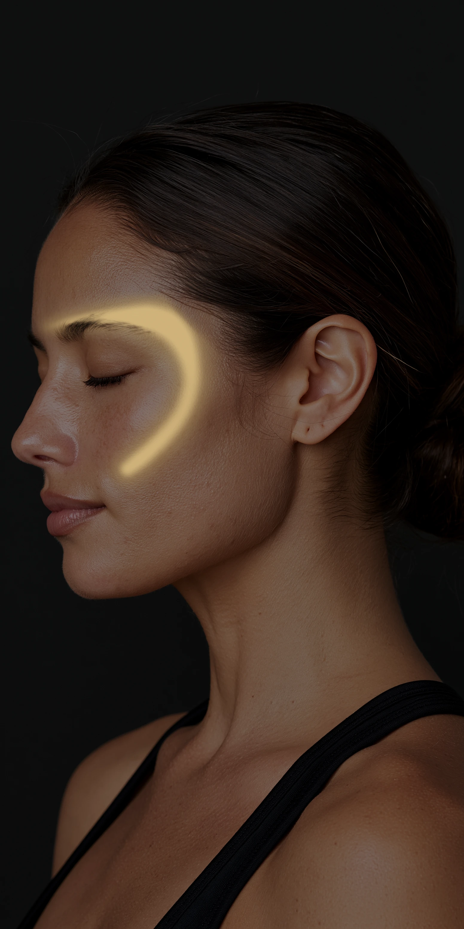
Where the pain is felt: The temple, the eyebrow, the upper teeth. Tooth pain with a healthy tooth is in the documented pattern.
Common causes: Jaw clenching, night grinding, gum chewing, long dental work, stress.
Patients say: Temple headaches. Teeth that ache with nothing wrong on the dental exam. A tired jaw by evening.
When to get evaluated: Jaw pain with chest pressure or shortness of breath during exertion is a cardiac evaluation, immediately. New temple pain over fifty with scalp tenderness or vision change is evaluated for temporal arteritis first.
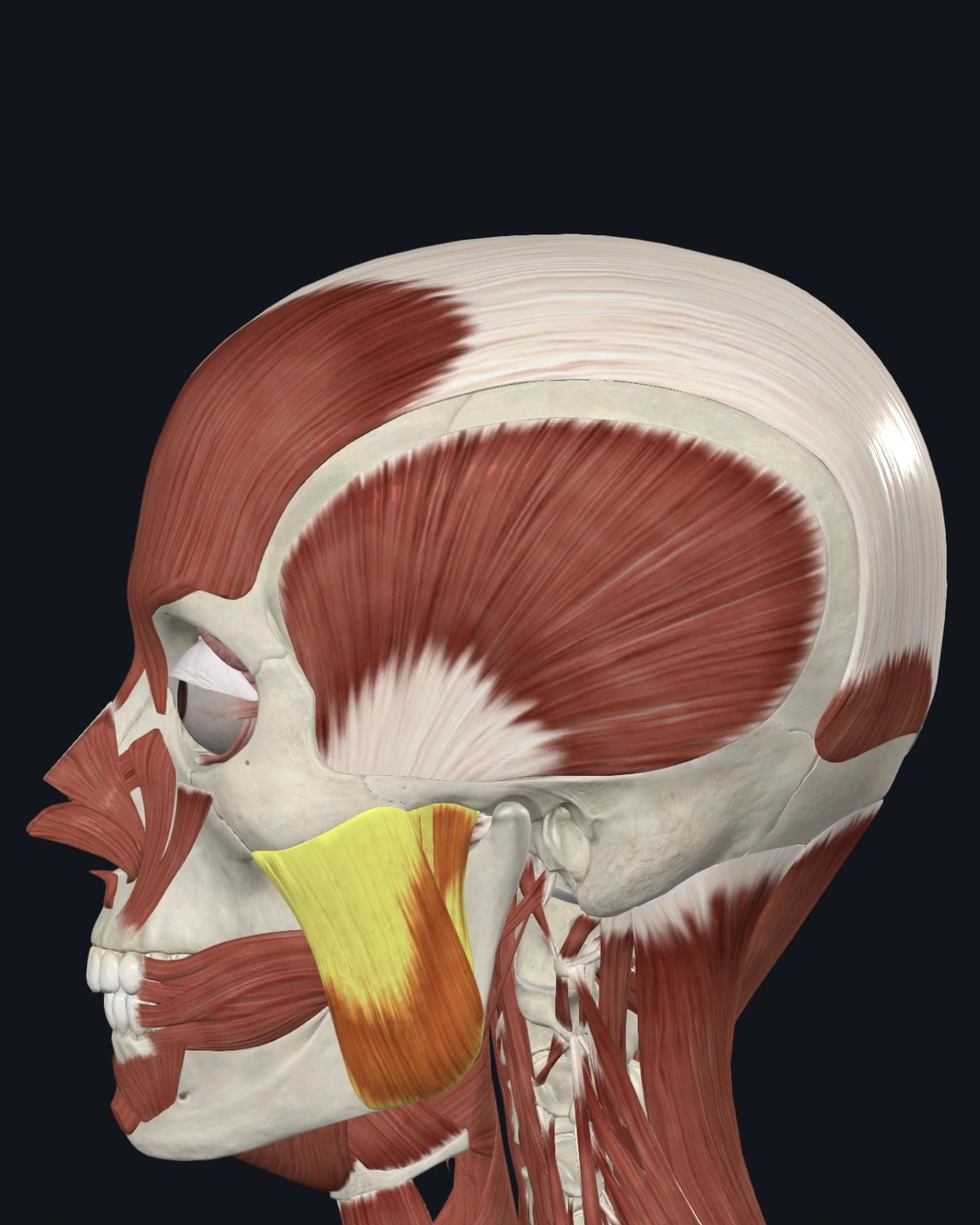
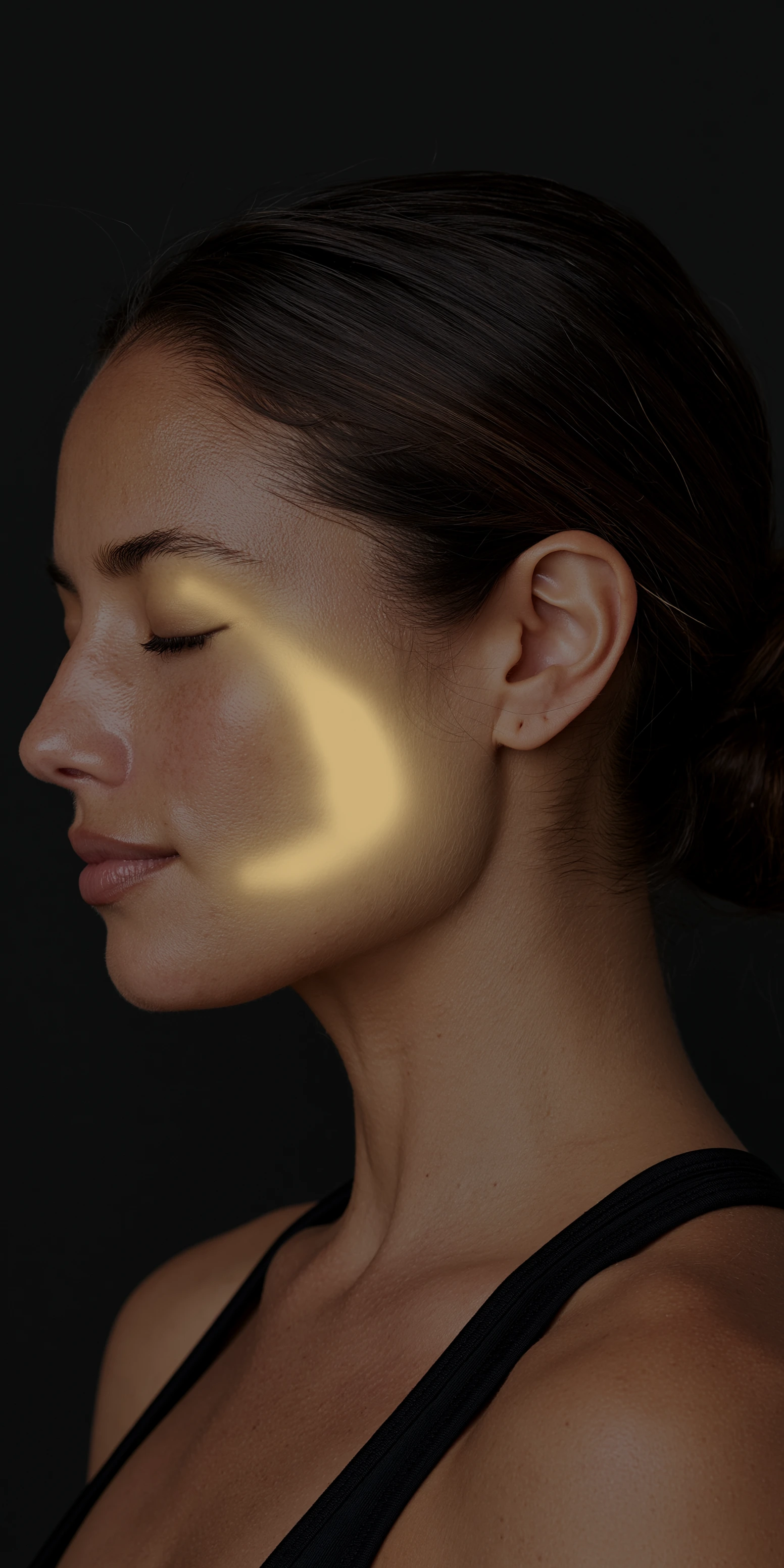
Where the pain is felt: The jaw, the ear, the lower teeth, and up to the eyebrow. Restriction shows as limited mouth opening.
Common causes: Clenching and grinding, chewing on one side, nail biting, jaw tension held under stress.
Patients say: Ear pain with a normal ear exam. Jaw fatigue when chewing. Clicking with tightness in the cheek.
When to get evaluated: A jaw that locks open or closed, or trauma to the jaw, is examined before treatment.
Shoulder & Upper Back
Six referral patterns are shown here.
The Muscle Library lists all fifteen patterns.
Each muscle shows where the pain is felt, what causes it, and how patients describe it.
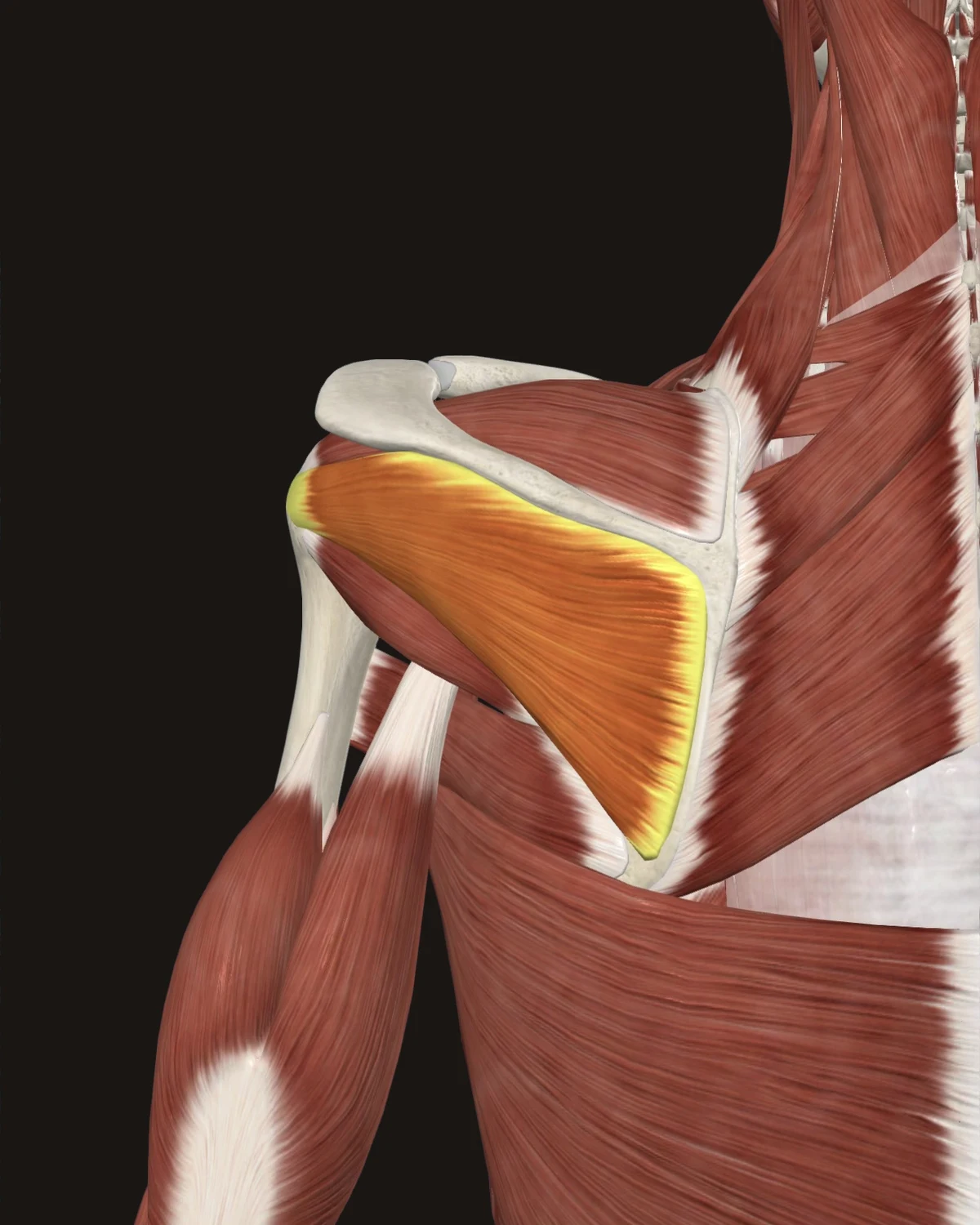
Where the pain is felt: Deep in the front of the shoulder, down the outside of the arm, into the upper back. The muscle sits on the shoulder blade; the pain sits in front. This mismatch is the region’s defining pattern.
Common causes: Reaching behind the body, throwing sports, swimming, repetitive overhead reaching, sleeping on the affected shoulder.
Patients say: “It hurts in the front, right here.” Trouble reaching into a back pocket or hooking a bra. Night pain lying on that side.
When to get evaluated: Inability to lift the arm after a fall, or a visible change in the shoulder’s shape, gets imaging before needling.
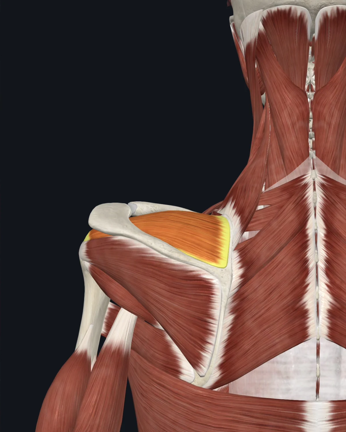
Where the pain is felt: The outer shoulder as a deep ache, down the outside of the arm toward the elbow. Aggravated by lifting the arm to the side.
Common causes: Carrying heavy loads with straight arms, overhead work, repetitive lifting, a sudden pull.
Patients say: A dull ache in the shoulder cap. Pain reaching to a high shelf. Trouble sleeping on that side.
When to get evaluated: Weakness raising the arm that does not vary with pain level suggests a structural tear. Evaluation first.
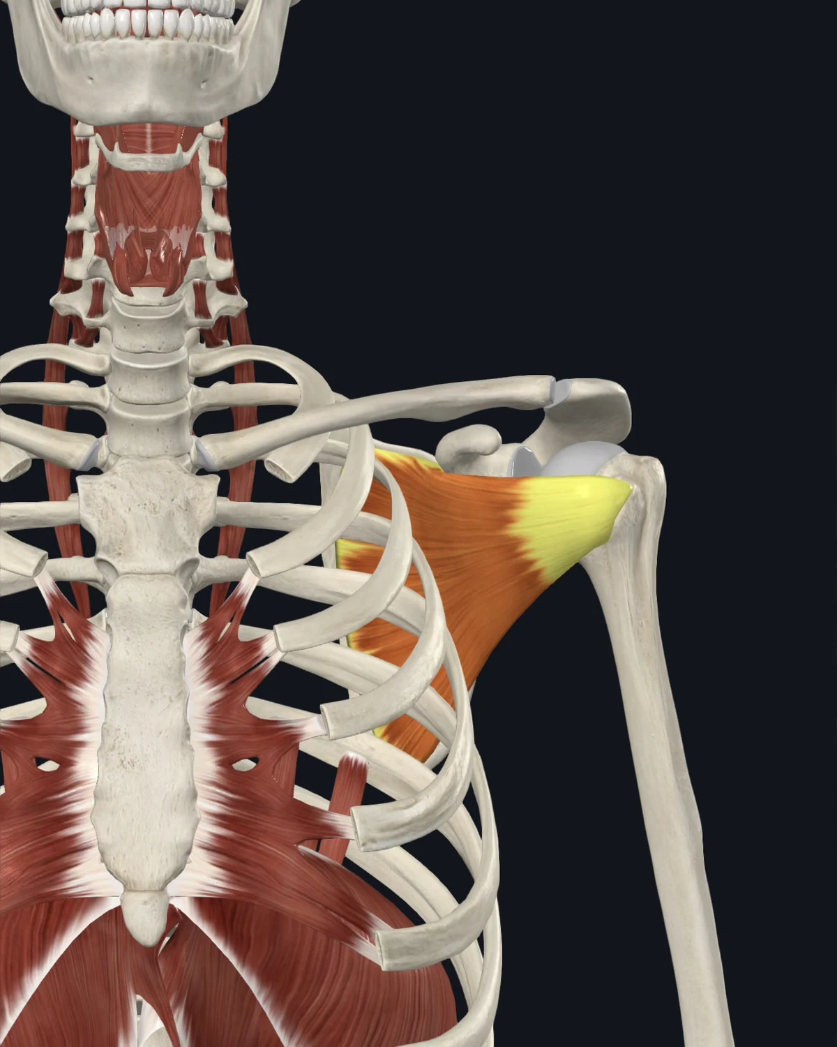
Where the pain is felt: The back of the shoulder, with a documented band of referral to the wrist. Severe restriction reads as frozen shoulder.
Common causes: Prolonged arm-at-side positioning after injury or surgery, swimming, throwing, protective disuse of the arm.
Patients say: “I can’t reach up or out.” A wrist band of ache with a normal wrist. Shoulder that keeps losing motion.
When to get evaluated: Progressive stiffness with night pain deserves a hands-on assessment to separate muscle restriction from true adhesive capsulitis.
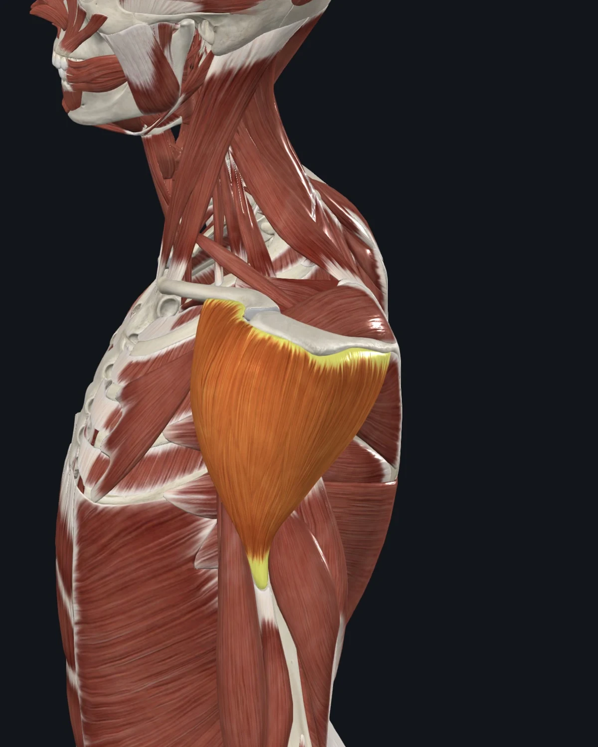
Where the pain is felt: Locally over the shoulder cap, front or back depending on the fibers involved. One of the maps where pain sits at its own location.
Common causes: Impact injuries, vaccination-site soreness that persists, overhead pressing, sudden loading.
Patients say: Soreness right where the shoulder rounds. Pain with lifting the arm in one specific direction.
When to get evaluated: Pain with numbness down the arm or into the hand brings the neck into the assessment, not just the shoulder.
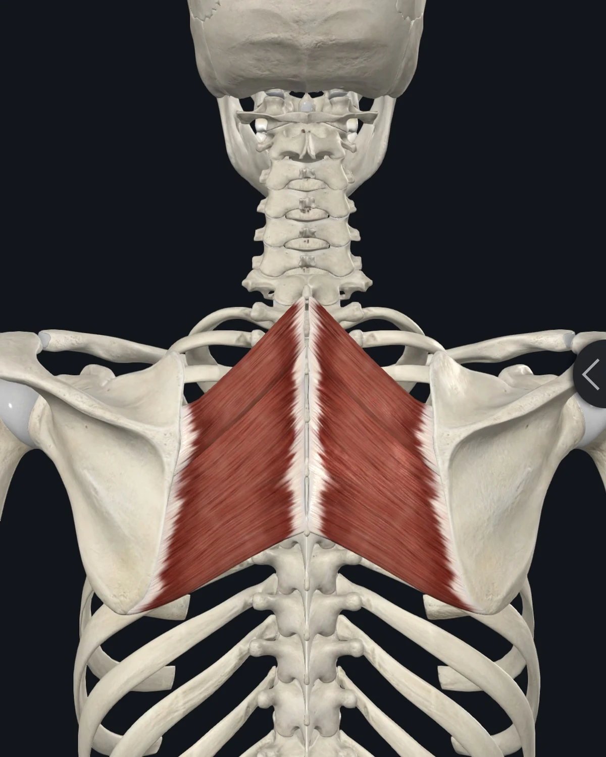
Where the pain is felt: Between the shoulder blade and the spine. A concentrated ache along the blade’s inner edge.
Common causes: Rounded-shoulder desk posture, long driving, carrying children, rowing and pulling exercise loads.
Patients say: “Right between my shoulder blade and spine, I can point to it.” The urge to press a thumb into the spot. Burning by the end of a work day.
When to get evaluated: Between-the-blades pain arriving with shortness of breath, chest pressure, or exertion needs cardiac evaluation before anything else.
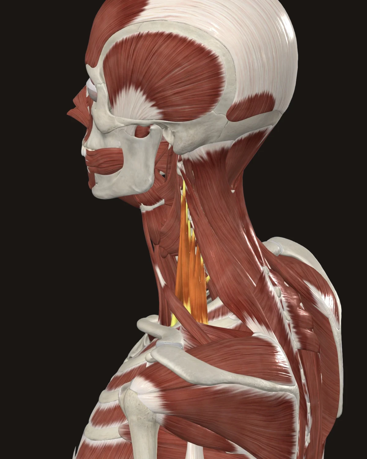
Where the pain is felt: The chest, the upper back, and down the arm into the thumb side of the hand. A neck muscle producing arm symptoms is the pattern’s signature.
Common causes: Chest breathing under stress, chronic cough, forward-head posture, carrying loads with straight arms.
Patients say: Arm heaviness or tingling that comes and goes. Hand symptoms without a clear wrist problem. Ache in the chest wall with normal cardiac workup.
When to get evaluated: Constant numbness, a weakening grip, or a cold pale hand involves nerve and vascular structures. Examination first.
Arm & Hand
Five referral patterns are shown here.
The Muscle Library lists all twenty-one patterns.
Each muscle shows where the pain is felt, what causes it, and how patients describe it.
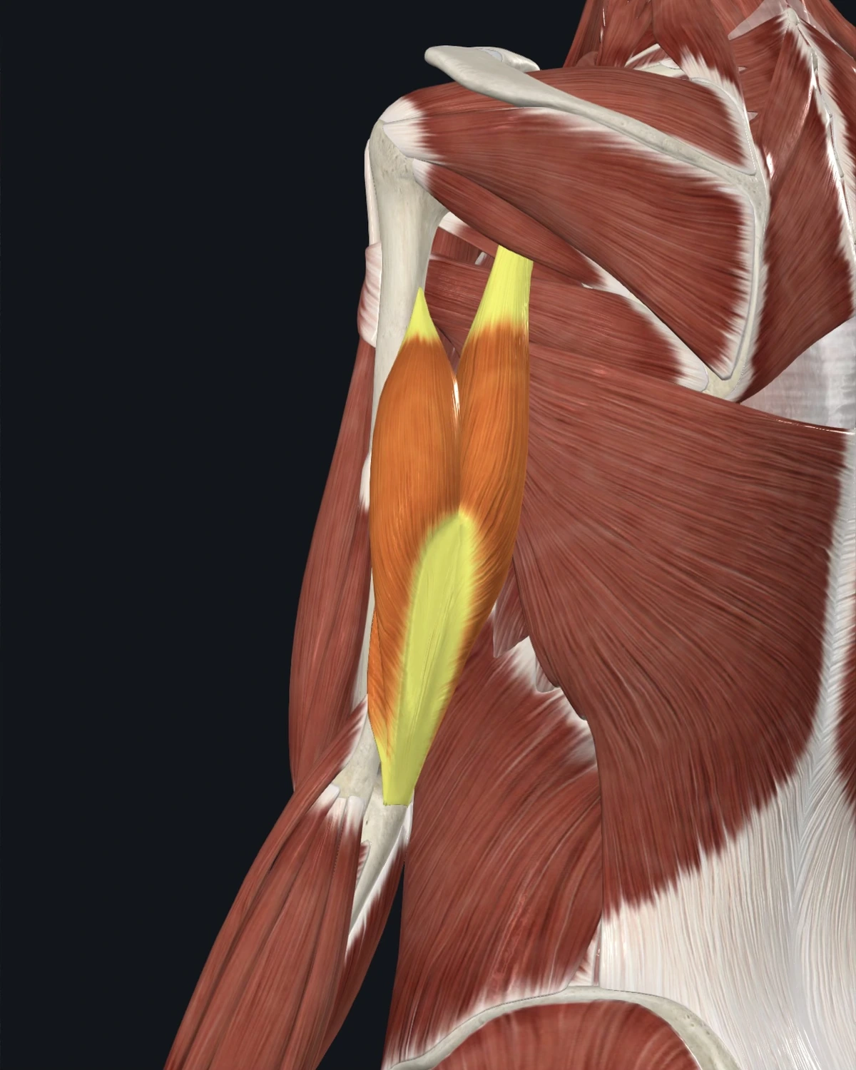
Where the pain is felt: The back of the arm, the outer elbow, and down to the ring and little fingers in the documented maps.
Common causes: Pushing exercise loads, prolonged leaning on elbows, racket sports, repetitive hammering or pressing.
Patients say: A deep ache in the back of the arm. Elbow soreness that moves. Pain pressing up from a chair.
When to get evaluated: Sudden weakness straightening the elbow after a forceful load gets assessed for tendon injury.
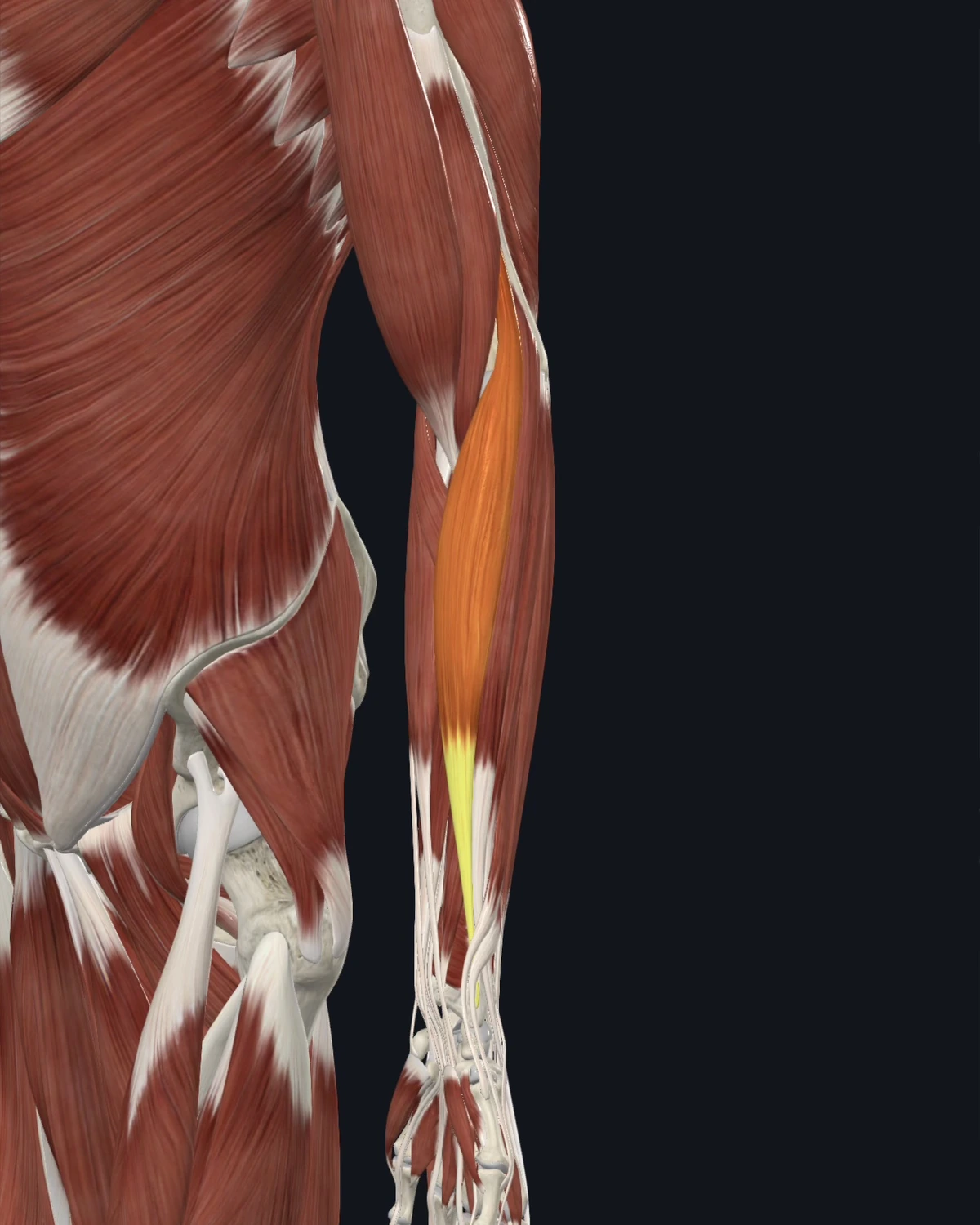
Where the pain is felt: The outer elbow, the top of the forearm, and the web of the thumb. Sits inside the “tennis elbow” complaint.
Common causes: Gripping with the thumb up, handshakes and tool use, carrying bags by the handles, racket and pulling sports.
Patients say: Pain at the outer elbow with gripping. An ache down the top of the forearm. Weak grip on bad days.
When to get evaluated: Elbow pain with locking or a joint that will not fully straighten is examined for the joint itself.
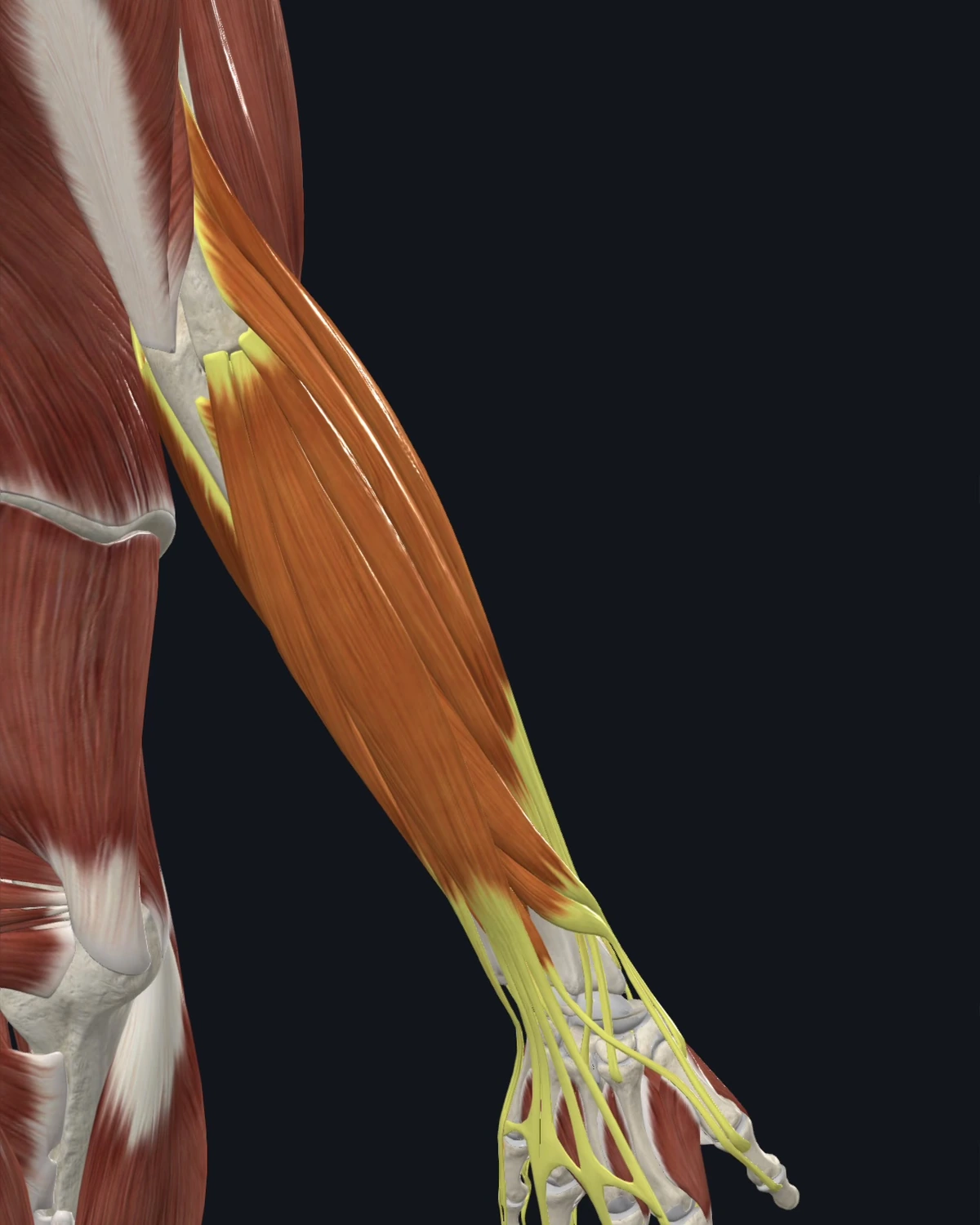
Where the pain is felt: The outer elbow and the back of the wrist and hand. The classic tennis-elbow pattern lives in these muscles.
Common causes: Keyboard and mouse hours, gripping tools, lifting with palms down, new racket or climbing loads.
Patients say: “Tennis elbow, but I don’t play tennis.” Wrist ache after a long computer day. Pain twisting a jar lid.
When to get evaluated: Numbness or tingling in specific fingers points at nerve involvement and changes the plan.
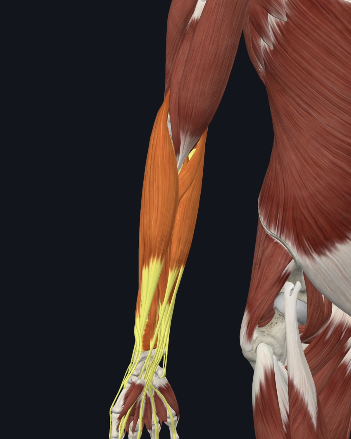
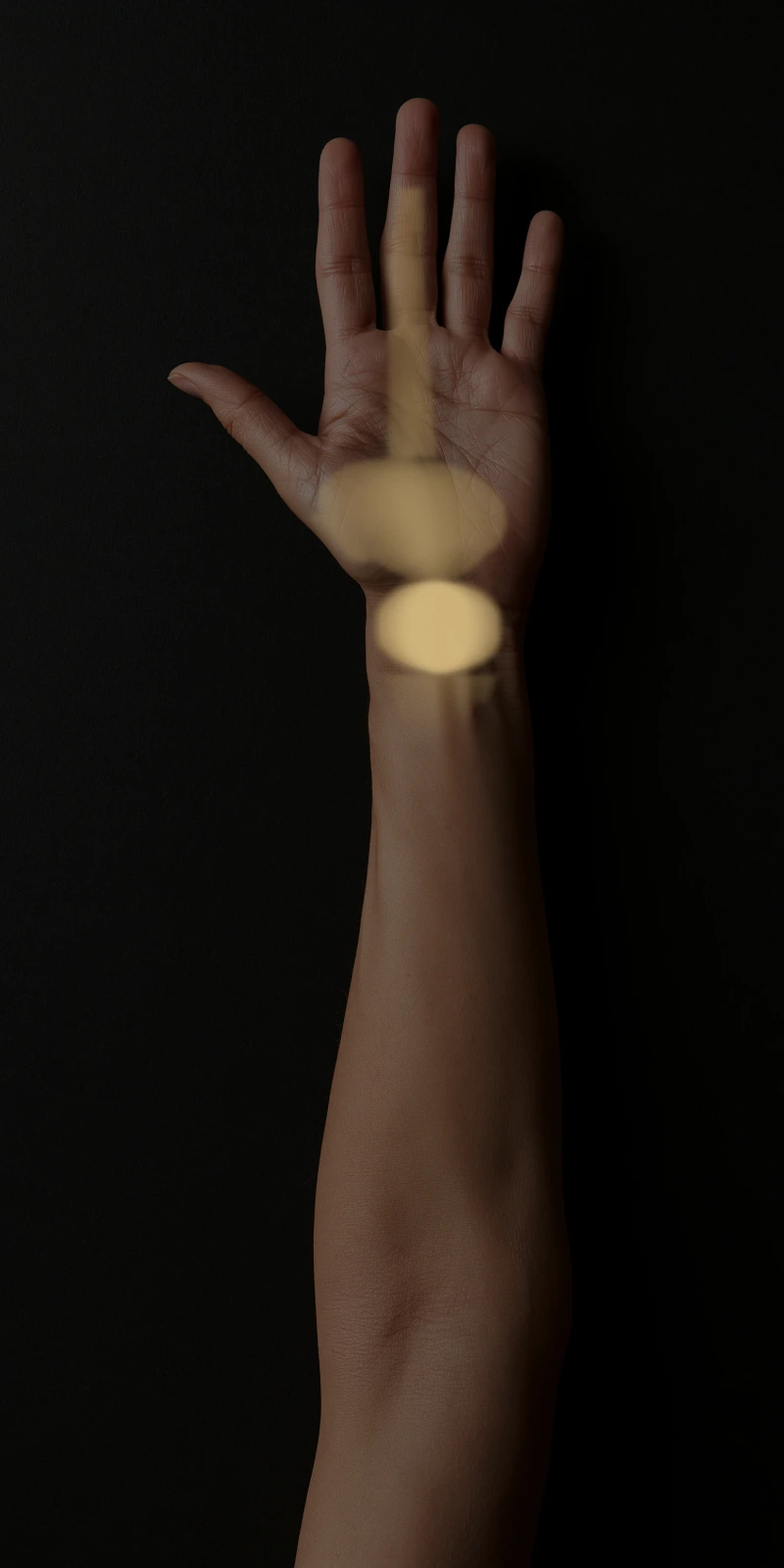
Where the pain is felt: The inner elbow and into the palm-side wrist and fingers. The golfer’s-elbow pattern.
Common causes: Gripping and curling loads, swinging clubs and bats, carrying with palms up, wrist-heavy work.
Patients say: Inner elbow soreness with gripping. Palm-side wrist ache. Fingers that feel tight in the morning.
When to get evaluated: Night tingling in the thumb, index, and middle fingers is assessed for carpal tunnel involvement before needling is assumed the answer.
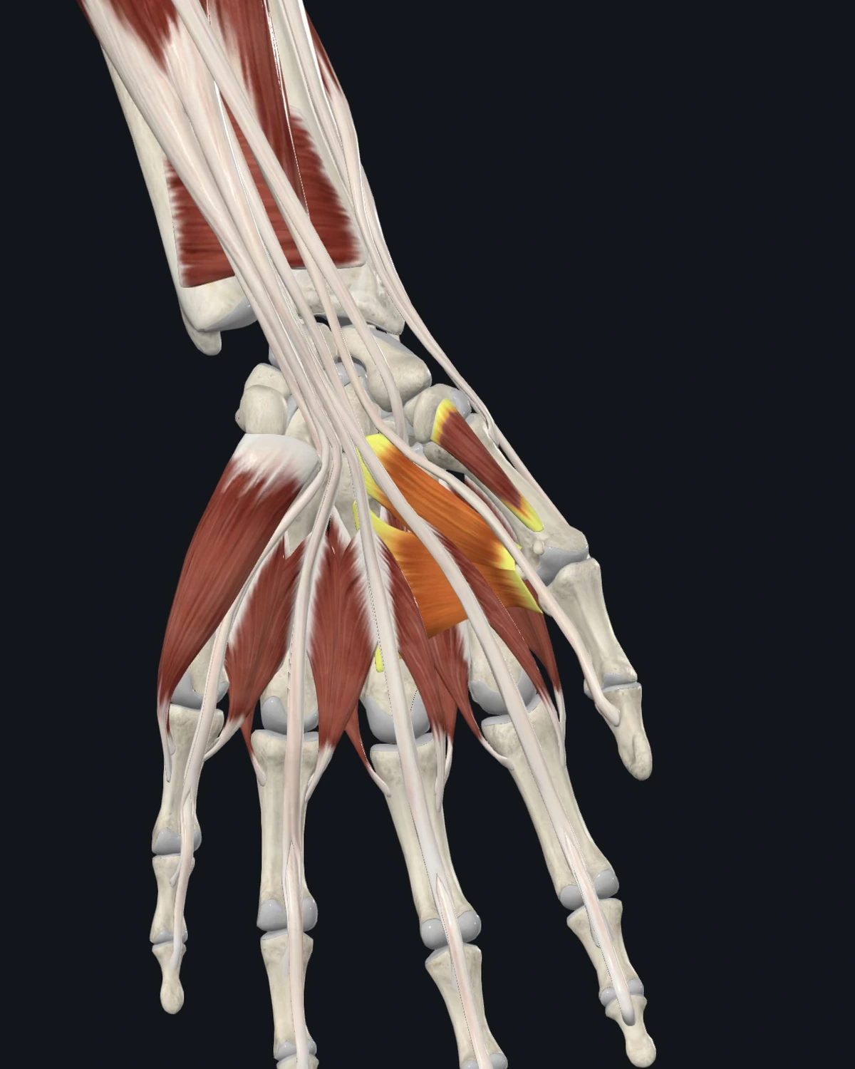
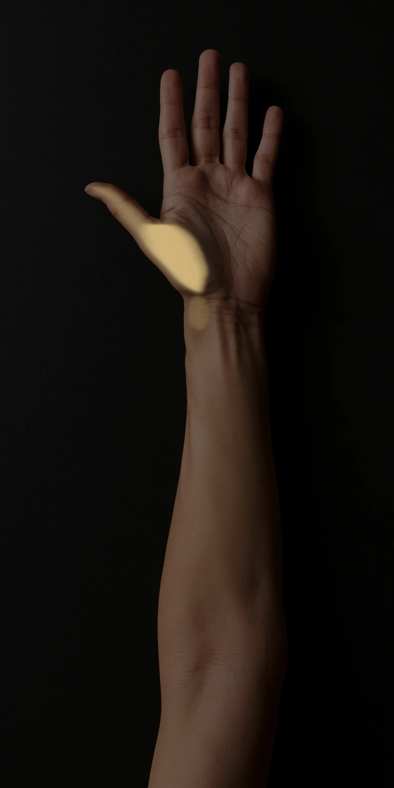
Where the pain is felt: The base of the thumb and the thumb side of the wrist. Aggravated by pinch and grip.
Common causes: Phone texting, gardening with hand tools, chopping, sustained pinch work, new parents lifting infants.
Patients say: “The base of my thumb aches with everything I grip.” Pain opening jars. Texting soreness.
When to get evaluated: A thumb base that is swollen, unstable, or painful at rest without use gets the joint examined for arthritis involvement.
Torso & Abdomen
Four referral patterns are shown here.
The Muscle Library lists all eleven patterns.
Each muscle shows where the pain is felt, what causes it, and how patients describe it.
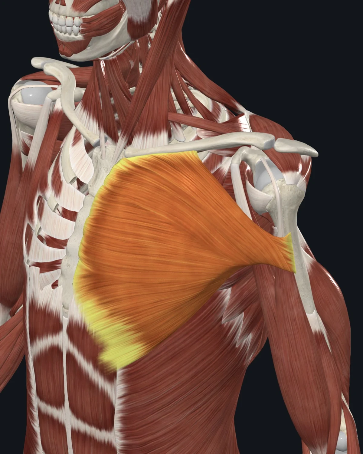
Where the pain is felt: The chest, the front of the shoulder, down the inner arm. The documented pattern overlaps the distribution people fear as cardiac.
Common causes: Rounded-forward posture, heavy pressing exercise, carrying loads against the chest, prolonged desk reach.
Patients say: Chest wall soreness that changes with arm movement. A tight line into the inner arm. Tenderness pressing the chest muscle itself.
When to get evaluated: This line is absolute: chest pain with exertion, pressure, shortness of breath, sweating, or nausea is treated as cardiac until a physician rules otherwise. Muscle explanations come after that, never before.
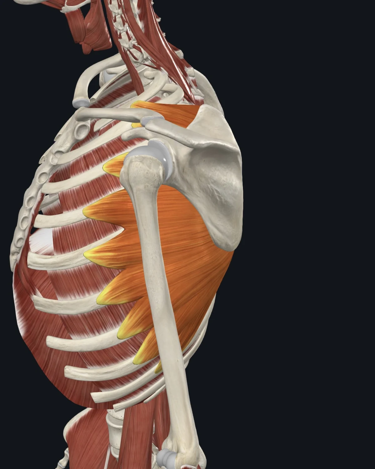
Where the pain is felt: The side of the ribs, with a documented reach to the inner border of the shoulder blade. A “stitch in the side” at rest fits the map.
Common causes: Hard running, chronic cough, heavy pressing, sustained twisting work.
Patients say: A side stitch that never fully leaves. Rib soreness taking a deep breath. An ache pinned to one spot on the ribs.
When to get evaluated: Rib pain after impact or a fall, or breathing pain with fever, is imaged and examined first.
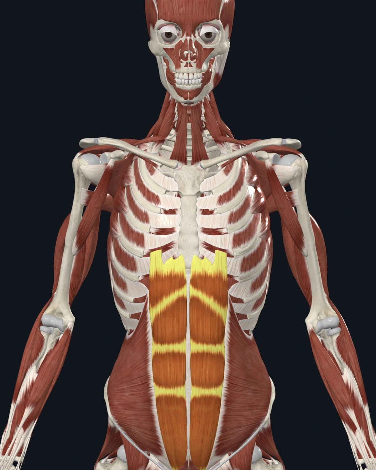
Where the pain is felt: Across the mid back at belt height in the documented maps, and locally through the abdomen. An abdominal muscle producing back pain is the pattern worth knowing.
Common causes: Sit-up-heavy training, chronic breath holding, surgical scarring, sustained slouched sitting.
Patients say: A band of back ache with a normal spine exam. Abdominal wall soreness with twisting. Cramping that tracks with muscle use, not meals.
When to get evaluated: Abdominal pain with fever, vomiting, changed bowel habits, blood, or weight loss is organ territory. Medical evaluation comes first, without exception.
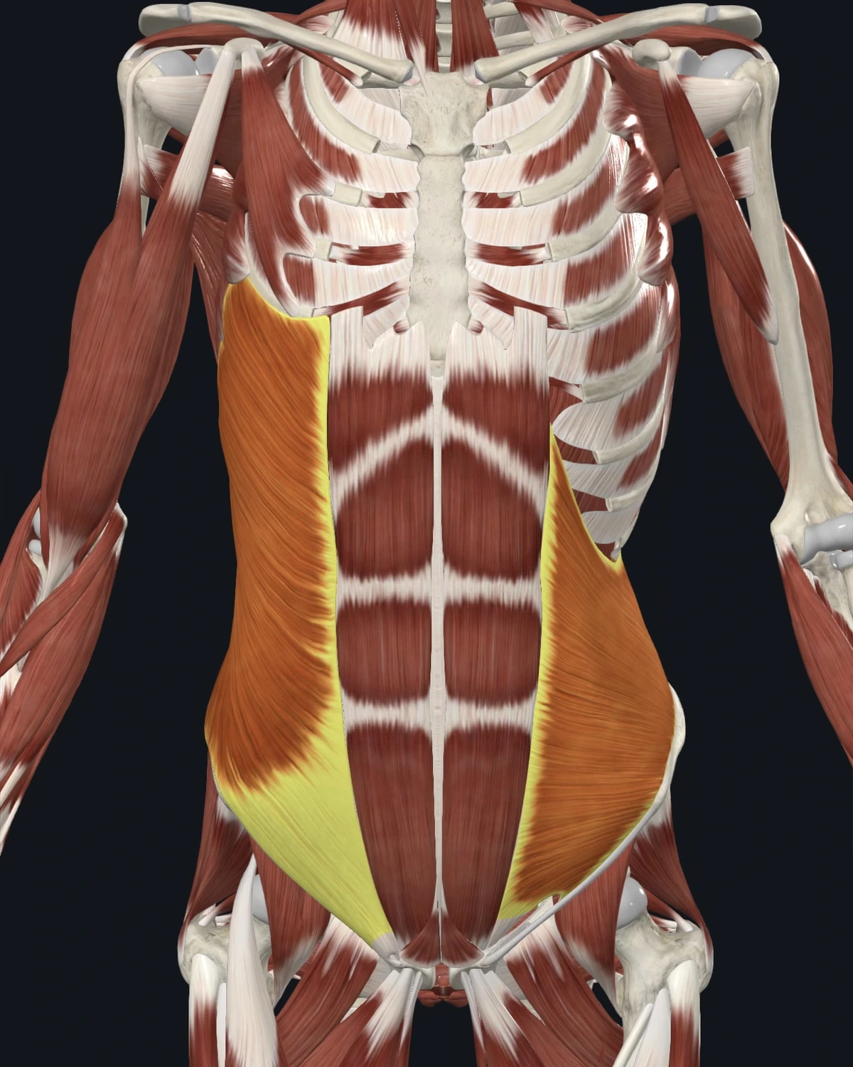
Where the pain is felt: Across the abdomen and into the groin in the documented maps, with heartburn-like symptoms recorded for upper fibers.
Common causes: Twisting sports, shoveling and raking, chronic cough, one-sided carrying.
Patients say: Groin ache with a normal hernia exam. Side soreness with rotation. A burning line that antacids never touch.
When to get evaluated: A visible bulge, groin pain with lifting that eases lying down, or any of the organ signs above gets medically assessed before muscle work.
Low Back & Hip
Six referral patterns are shown here.
The Muscle Library lists all fifteen patterns.
Each muscle shows where the pain is felt, what causes it, and how patients describe it.
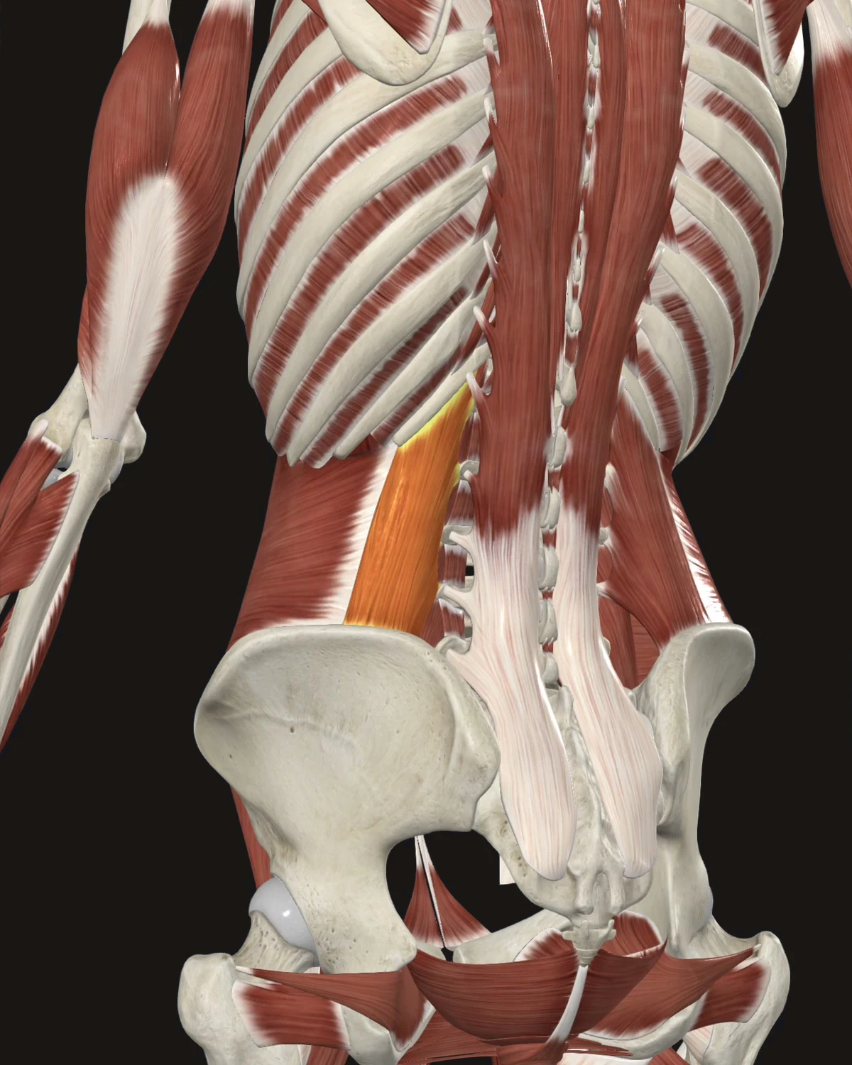
Where the pain is felt: The sacroiliac region, lower buttock, and along the iliac crest; the pattern extends toward the groin in the documented maps. Felt as deep, aching low back pain, aggravated by standing and by rolling over in bed.
Common causes: Sustained leaning postures, lifting combined with twisting, leg-length difference, one-sided carrying, gardening, shoveling.
Patients say: “It grabs when I stand up.” Difficulty rolling over at night. Pain on coughing or sneezing. Leaning away from the painful side.
When to get evaluated: Constant night pain unrelieved by position change, numbness, or bowel/bladder changes are not muscle-pattern findings. Those need medical evaluation.
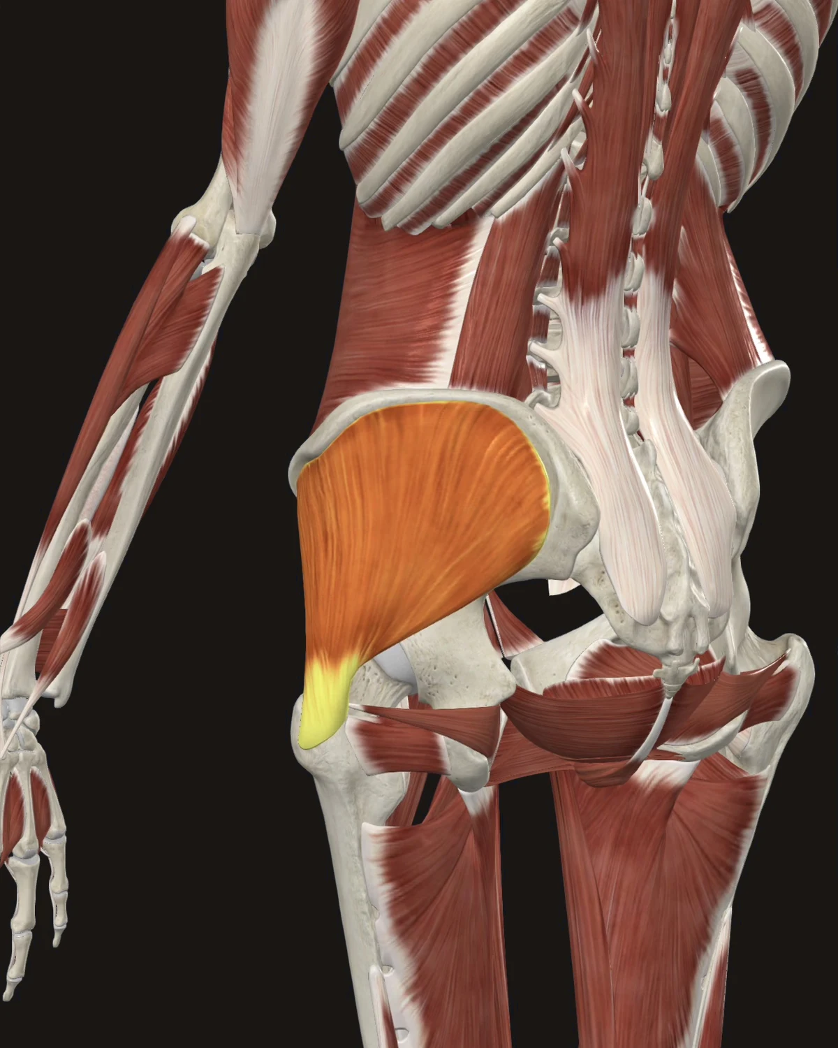
Where the pain is felt: The posterior iliac crest, the sacrum, and the outer buttock, extending into the upper posterior thigh. Read by patients as low back pain.
Common causes: Prolonged standing, walking on soft or uneven ground, rapid increases in running volume, standing with weight on one leg, a fall onto the hip.
Patients say: Aching over the belt line. Pain lying on the affected side at night. Soreness after long walks.
When to get evaluated: Weight-bearing pain that began with a fall gets imaging before needling.
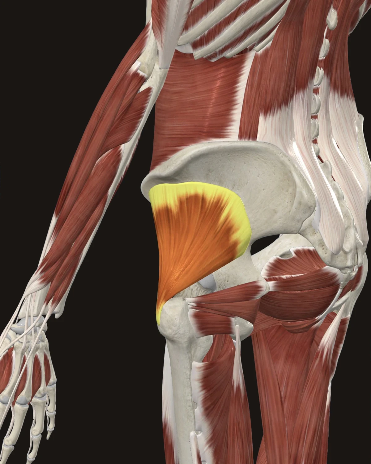
Where the pain is felt: Down the lateral or posterior thigh into the calf. The mapped pattern reaches the ankle without nerve involvement. This is the classic sciatica mimic.
Common causes: Sudden overload (a misstep off a curb), long drives, sitting on a wallet, compensation for a painful hip.
Patients say: “Sciatica” that does not behave like nerve pain: no true numbness, worst on standing up from sitting, walking helps and then worsens it.
When to get evaluated: True numbness, foot weakness, or reflex changes indicate nerve involvement. The first-visit assessment separates the two.
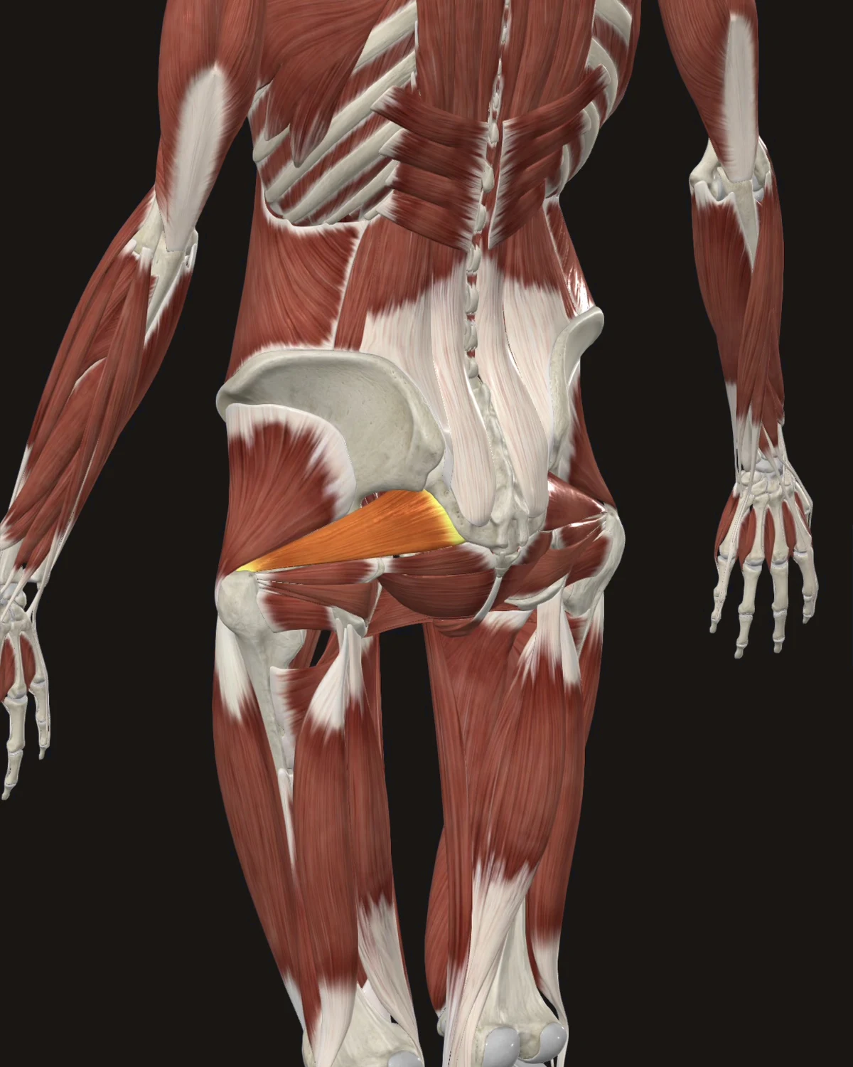
Where the pain is felt: The sacroiliac region, the buttock, and the posterior thigh. The sciatic nerve passes beside this muscle, and in some people through it, so piriformis tightness can also produce true nerve irritation.
Common causes: Prolonged sitting, driving, running on sloped road surfaces, repeated hip external rotation in sport.
Patients say: Deep buttock ache, worst with sitting, sometimes with leg symptoms. Discomfort getting in and out of the car.
When to get evaluated: Neurological signs move this from pattern education to hands-on examination.
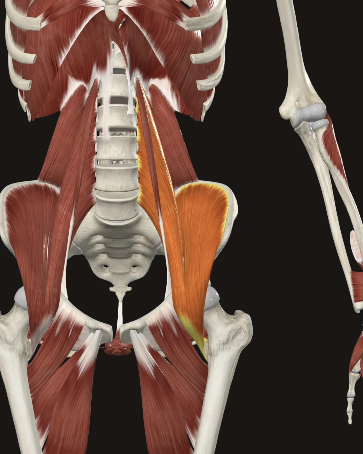
Where the pain is felt: Vertically along the lumbar spine, and to the front of the hip and upper thigh. The signature presentation: low back pain that eases lying with the knees bent.
Common causes: Prolonged sitting at a desk or wheel, sit-up-heavy training, sleeping curled, sprinting after a period of inactivity.
Patients say: Trouble standing fully upright after sitting. “My back is tight until I get moving.” A pinch at the front of the hip on stairs.
When to get evaluated: Groin pain with clicking, locking, or giving way points to the joint or labrum, not muscle. Assessment first.
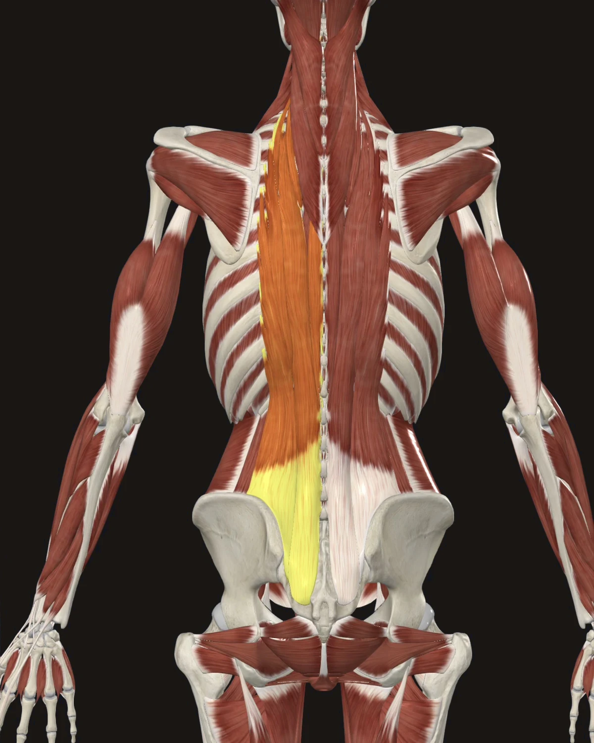
Where the pain is felt: Locally along the spine and downward toward the buttock.
Common causes: Lifting in a rounded-back position, long unsupported sitting, deconditioning, protective muscle tension held after an acute episode.
Patients say: Broad stiffness. “A band across my back.” Morning stiffness that eases with movement.
When to get evaluated: Fever, unexplained weight loss, or pain following trauma are screening findings addressed before any needling plan.
Thigh & Knee
Five referral patterns are shown here.
The Muscle Library lists all fourteen patterns.
Each muscle shows where the pain is felt, what causes it, and how patients describe it.
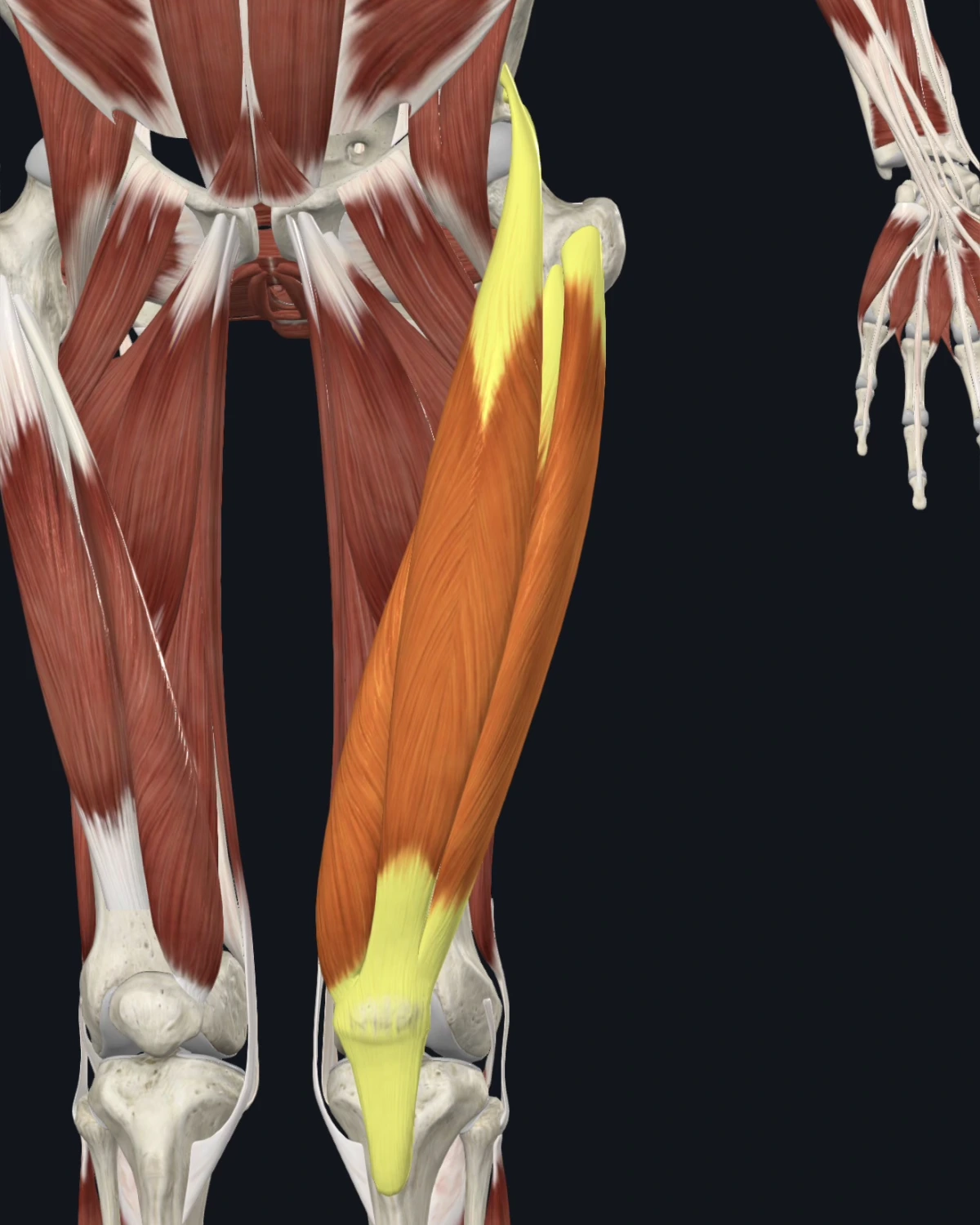
Where the pain is felt: The front of the knee and deep in the kneecap region, with the rectus femoris pattern reaching down from the hip. Knee pain from thigh muscle is this region’s core lesson.
Common causes: Hill and stair loads, squatting volume, direct blows to the thigh, sudden sprinting, long sitting that shortens the hip.
Patients say: “My knee hurts but the scans are clean.” Kneecap ache on stairs, worse going down. A knee that buckles without swelling.
When to get evaluated: A knee that locks, gives way with swelling, or swelled within hours of an injury is examined for the joint itself first.
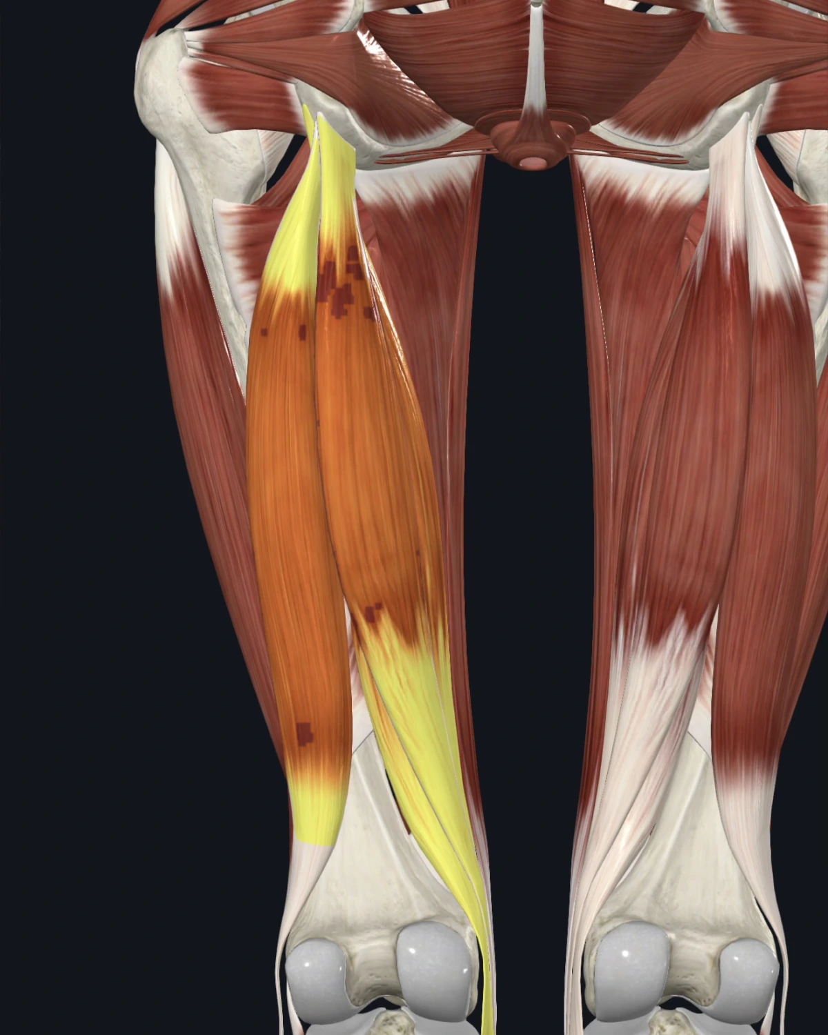
Where the pain is felt: The back of the thigh up to the lower buttock and down to the back of the knee. Sitting pressure aggravates it in the documented pattern.
Common causes: Sprinting and sudden acceleration, long sitting on hard edges, insufficient recovery between training loads, compensation for hip weakness.
Patients say: An ache sitting on hard chairs. Back-of-knee tightness. A pull that never fully healed.
When to get evaluated: A pop with immediate bruising and weakness is assessed for a true tear before needling.
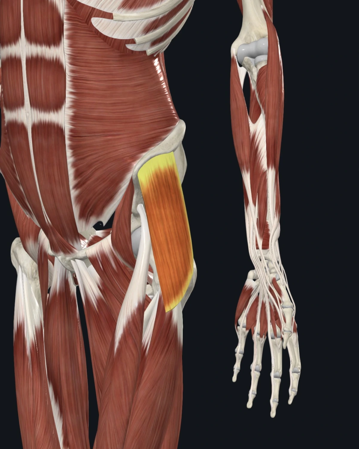
Where the pain is felt: The outer hip and down the outer thigh toward the knee. Lies inside the “IT band pain” complaint.
Common causes: Running volume increases, cambered surfaces, standing with weight shifted to one hip, weak gluteal support.
Patients say: Outer hip ache lying on that side. A line of tightness down the outside of the thigh. Runner’s outer-knee pain.
When to get evaluated: Outer hip pain with true weakness lifting the leg sideways, or pain after a fall in an older adult, is imaged first.
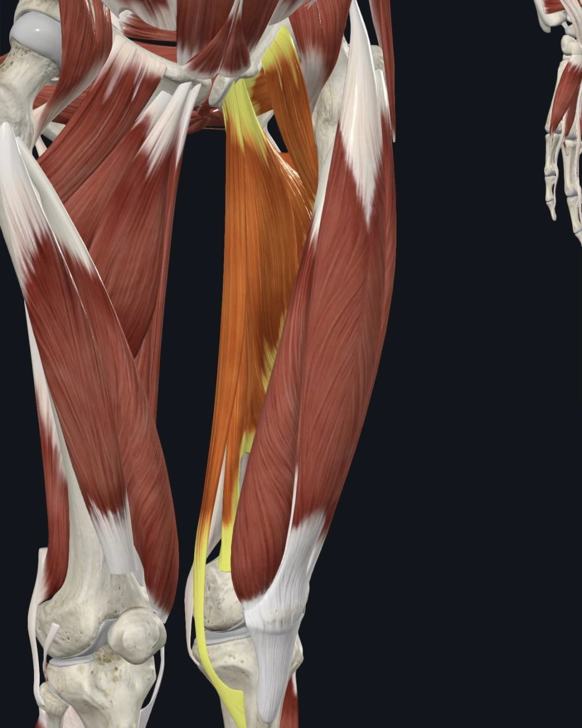
Where the pain is felt: The groin and inner thigh, reaching to the inner knee in the documented maps.
Common causes: Skating and cutting sports, sudden side lunges, riding, rapid return to sport after time off.
Patients say: Groin pull that keeps returning. Inner knee ache with a clean knee exam. Pain pushing off sideways.
When to get evaluated: Groin pain with a bulge, with coughing, or unrelated to movement is examined for hernia and hip joint involvement.
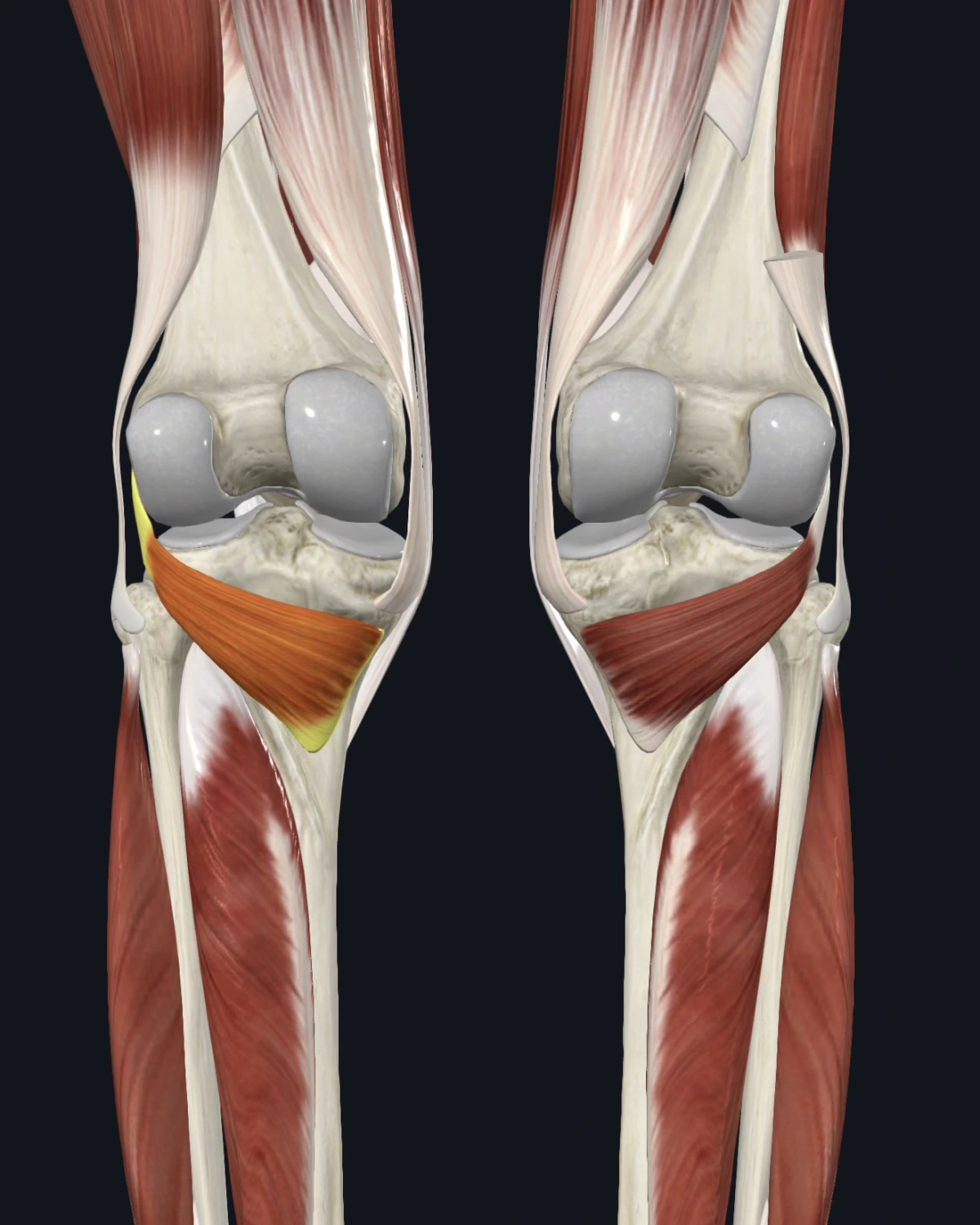
Where the pain is felt: The back of the knee. A small muscle with a precise, local map.
Common causes: Downhill walking and running, sudden twisting on a planted foot, unstable footwear.
Patients say: “Right behind my knee, especially downhill.” Stiffness at the back of the knee crouching. Pain at push-off.
When to get evaluated: Back-of-knee swelling or a calf that is swollen, warm, or tender raises circulation questions answered by a clinician, urgently.
Leg, Ankle & Foot
Five referral patterns are shown here.
The Muscle Library lists all twenty patterns.
Each muscle shows where the pain is felt, what causes it, and how patients describe it.
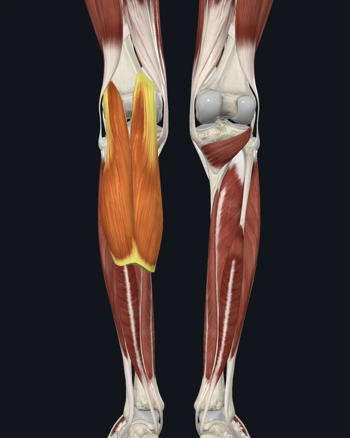
Where the pain is felt: The calf itself and the instep of the foot. Night calf cramps sit in this muscle’s documented territory.
Common causes: Hill work, high heels and stiff dress shoes, cold-weather running, dehydration, long drives with a pointed foot.
Patients say: Instep ache after runs. Calf cramps that strike at night. Tightness that never stretches out.
When to get evaluated: One calf that is swollen, warm, red, or tender without injury is evaluated for a clot the same day. This is not a wait-and-see finding.
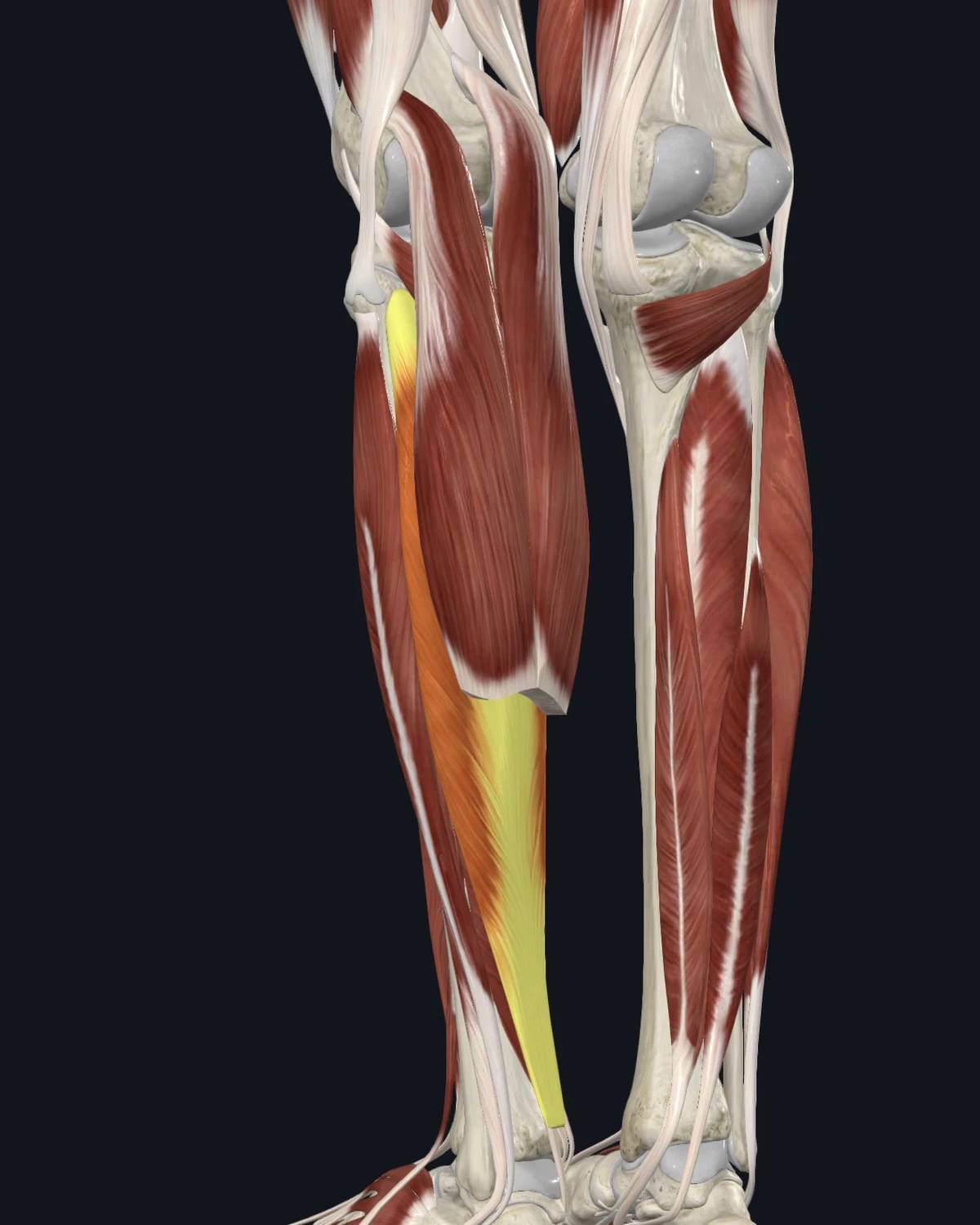
Where the pain is felt: The heel and the back of the ankle, with the documented pattern reaching the sacroiliac region. Heel pain labeled plantar fasciitis sits partly in this map.
Common causes: Running volume, calf-dominant sports, footwear changes to flat shoes, prolonged tiptoe work.
Patients say: Heel pain with the first steps of the morning. An ache up the Achilles line. Calf fatigue climbing stairs.
When to get evaluated: A sudden snap in the calf or heel with immediate weakness is assessed for tendon rupture before anything else.
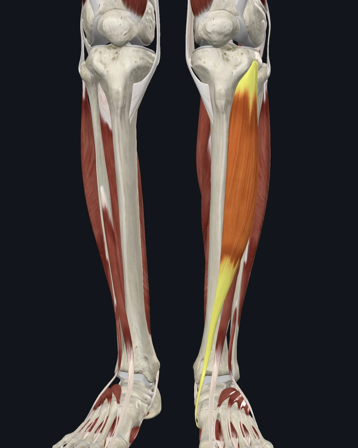
Where the pain is felt: The front of the ankle and the big toe. The documented line runs down the shin into the foot.
Common causes: New or increased running, downhill loads, tight calves forcing extra lift work, hard-surface walking in flat shoes.
Patients say: “Shin splints” along the front. Ankle ache with a clean ankle exam. A big toe that hurts to lift.
When to get evaluated: Shin pain that is focal on the bone, worse with every impact, and present at rest is assessed for stress fracture.
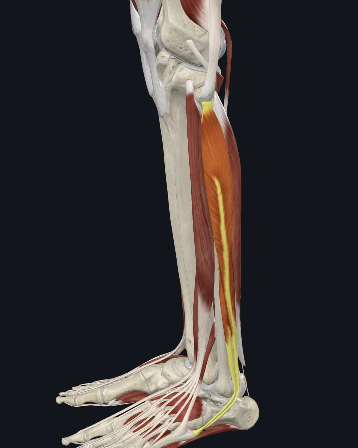
Where the pain is felt: The outer ankle, behind and below the ankle bone. The pattern imitates a sprain that never healed.
Common causes: Old ankle sprains, walking on uneven ground, unstable footwear, weak lateral hip support up the chain.
Patients say: An outer ankle that aches long after the sprain healed. A “weak” ankle that rolls easily. Soreness after uneven trails.
When to get evaluated: An ankle that gives way with true instability on exam, or pain with inability to bear weight after a twist, is imaged first.
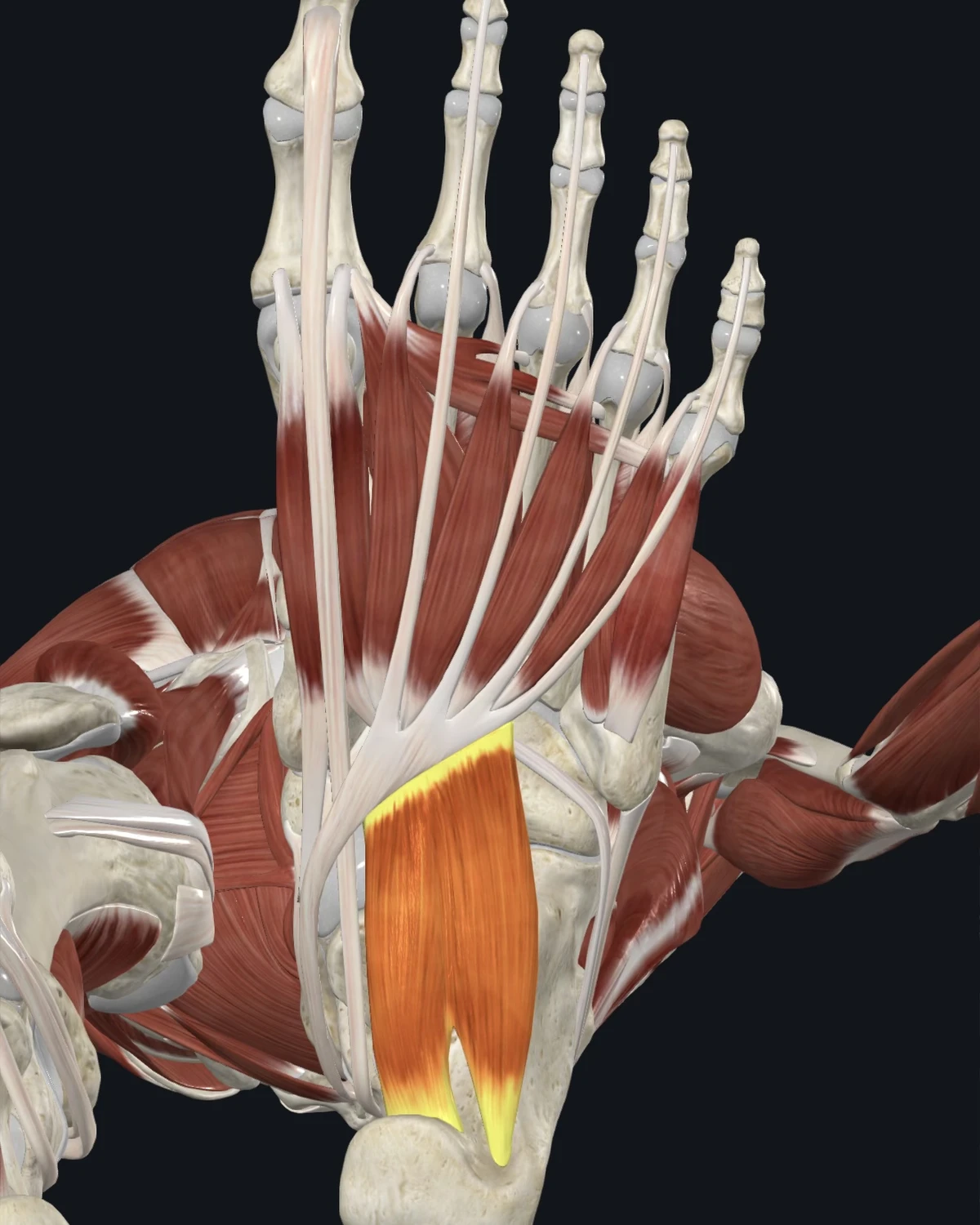
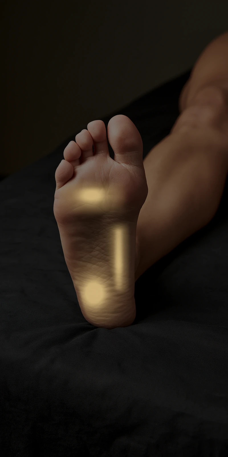
Where the pain is felt: The sole of the foot, the heel, and the ball of the foot. Together with the calf maps, this is where persistent plantar heel pain lives.
Common causes: Unsupportive footwear, sudden barefoot volume, standing occupations, high-arch and flat-foot mechanics.
Patients say: Arch ache by afternoon. Ball-of-foot burning in thin shoes. Heel pain that moved in and stayed.
When to get evaluated: Numbness or burning in a defined band of the sole is assessed for nerve entrapment. Foot pain with fever or in a person with diabetes goes to medical evaluation first.